Neurochirurgie
La Neurochirurgie, d'hier à aujourd'hui.
Les origines de la Neurochirurgie
L’homme a toujours été intéressé par ce qu’il se passe dans sa tête. Dès le Néolithique, la trépanation s’inscrit comme la première trace d’intervention physique sur le crâne, même si ce n’était pas principalement pour des raisons médicales. Il faudra attendre le XIXe siècle et les découvertes en anatomie cérébrale et en anesthésie pour que naisse réellement la Neurochirurgie. En 1887, Gowers et Horsley cherchent à rendre les opérations moins risquées. En effet à cette époque, peu de patients survivaient pendant ou à la suite d’une intervention. Il n’existait aucun moyen efficace et sans danger de réaliser une hémostase. Ils développent alors l’utilisation de « cautères galvaniques », une simple boucle de fil chauffée. Après plusieurs tentatives, les résultats n’étaient pas satisfaisants et l’intérêt médical pour la neurochirurgie déclina car trop risquée. En 1902, Harvey Cushing est le premier chirurgien américain à opérer une tumeur du cerveau avec succès. Il propose également l’utilisation de rayon X pour le diagnostic de tumeurs cérébrales, utilise des stimuli électriques pour étudier le cortex cérébral et participe, par exemple, au développement du bistouri électrique. En 1920, Walter Dandy son successeur publie son travail sur la production de liquide céphalo-rachidien. A l’aise avec les opérations du cerveau et de la moelle épinière, il devient le chirurgien le plus connu de son époque. Il décrit en 1921 une opération d’élimination des tumeurs de la région pinéale, en 1922 une élimination complète des tumeurs de l’angle cérébellopontine et l’utilisation de l’endoscopie pour le traitement de l’hydrocéphalie, en 1929 la suppression d’une hernie discale dans la colonne vertébrale et bien d’autres avancées encore. Depuis, de nombreux progrès en Neurochirurgie ont été induits par l’évolution des outils et instruments chirurgicaux comme la curette, le burin ou les crochets.
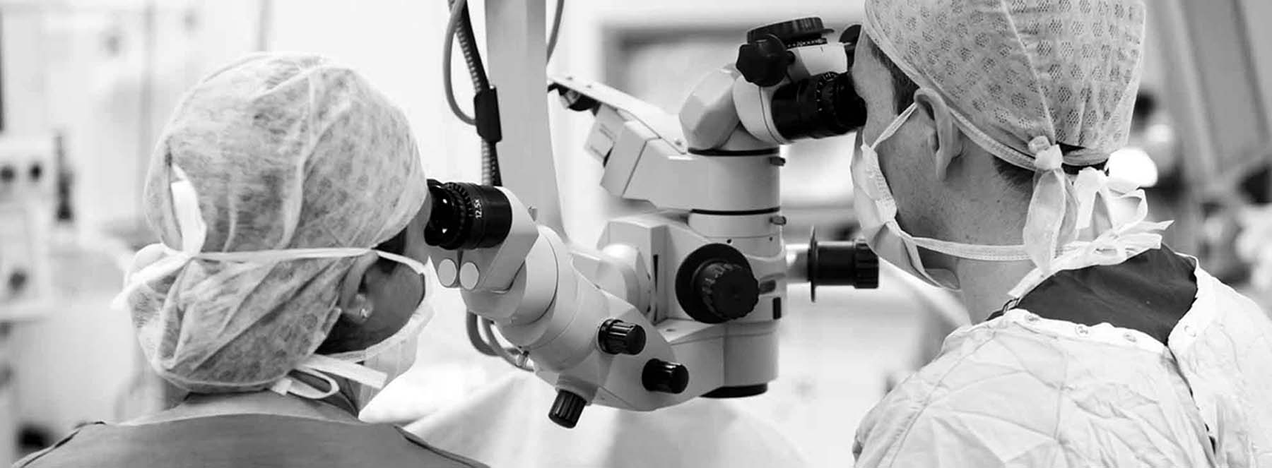
La Neurochirurgie moderne et nos solutions d’accompagnement
Aujourd’hui bien plus sûre et évoluée, la Neurochirurgie est une discipline médicale reconnue. Nos simulateurs sont des outils pédagogiques qui accompagnent l’enseignement et l’entraînement des apprenants que ce soit en formation initiale ou continue. Réalisme, précision et fidélité anatomique font de nos simulateurs de véritables atouts pour accompagner et accélérer la courbe d’apprentissage en Neurochirurgie.